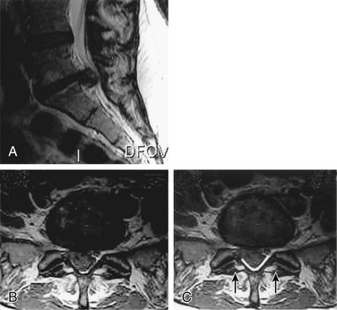
Magnetic resonance imaging mri is the imaging study of choice for the evaluation of suspected patients with ces due to its ability to accurately depict soft tissue pathology. Consequently the majority of urgent scans requested are normal.
Cauda Equina Syndrome An Overview Sciencedirect Topics
cauda equina syndrome with normal mri imaging
cauda equina syndrome with normal mri imaging is a summary of the best information with HD images sourced from all the most popular websites in the world. You can access all contents by clicking the download button. If want a higher resolution you can find it on Google Images.
Note: Copyright of all images in cauda equina syndrome with normal mri imaging content depends on the source site. We hope you do not use it for commercial purposes.
Cauda equina syndrome is a rare emergency with devastating consequences early recognition is paramount as the presence of bladder dysfunction portends bad functional outcomes the presence of bilateral lower extremity weakness or sensory changes should alert clinicians to the diagnosis.

Cauda equina syndrome with normal mri imaging. A large number of patients do not have cauda equina syndrome ces on mri to account for their clinical findings. Cauda equina compression mri this is an example of one of the most common indications for an emergency mri. Patients with complete cauda equina syndrome have a poorer outcome 3.
Such root dysfunction can cause a combination of clinical features but the term cauda equina syndrome is used only when these include impairment of bladder bowel or sexual. Cauda equina syndrome ces. We report a case of cauda equina syndrome caused by gnathostoma spinigerum which was confirmed by an immunoblotting test.
The cauda equina is formed by the nerve roots caudal to the level of the conus medullaris. This woman in her 40s presented with acute onset of lower limb weakness and urinary incontinence and her ed physician suspected compression of the caudal equina based on his clinical examination. The conus medullaris was slightly enlarged with abnormal enhancement.
While magnetic resonance imaging mri is used as the diagnostic gold standard for cauda equina syndrome ces many mri scans obtained from patients presenting with signs andor. Mr imaging of the lumbosacral spine showed long segmented hyperintensity along the cauda equina with irregular enhancement on the postcontrast study. The most common etiology of ces is a large central lumbar disc herniation at the l4 5 or l5 s1 level.
Cauda equina syndrome results from the dysfunction of multiple sacral and lumbar nerve roots in the lumbar vertebral canal. The nerves in the cauda equina region include the lower lumbar and all the sacral nerve roots. Chronic but surgical decompression within 24 hours seems to have the best outcomes 136.
We aimed to determine whether any clinical manifestation of ces as stated in royal college of radiology guidelines could predict the presence of established ces on mri. The cauda equina syndrome can result from any lesion that compresses the cauda equina and causes a dysfunction of multiple lumbar and sacral nerve roots. Consequently the majority of urgent scans requested are normal.
Cauda equina syndrome is considered a diagnostic and surgical emergency although there is some debate about the timing of surgery and depends on acute vs. The patient was treated with. A large number of patients do not have cauda equina syndrome ces on mri to account for their clinical findings.
Mr Imaging Findings In Cauda Equina Gnathostomiasis American
Cauda Equina Compression Mri Radiology At St Vincent S
Doing More With Less Diagnostic Accuracy Of Ct In Suspected Cauda
Lumbar Magnetic Resonance Imaging Mri Results Of A 64 Year Old
Editing Cauda Equina Syndrome Physiopedia
Cauda Equina Syndrome Spine Surgeon Vail Aspen Denver Co
Figure 1 From Cauda Equina Syndrome Following A Lumbar Puncture
Cauda Equina Syndrome Radiology Reference Article Radiopaedia Org
